Science Forum: Vision, challenges and opportunities for a Plant Cell Atlas
- Plant Cell Atlas, United States
- Department of Plant Biology, Carnegie Institution for Science, United States
- Department of Botany and Plant Sciences, University of California, Riverside, United States
- Joint Genome Institute, Lawrence Berkeley National Laboratory, United States
- Donald Danforth Plant Science Center, United States
- National Center for Genome Resources, United States
- Center for Genomics and Systems Biology, New York University, United States
- Department of Cell and Systems Biology, University of Toronto, Canada
- Department of Agronomy and Horticulture, University of Nebraska-Lincoln, United States
- Department of Cell and Systems Biology and the Centre for the Analysis of Genome Evolution and Function, University of Toronto, Canada
- Consejo Nacional de Investigaciones Científicas y Técnicas, Instituto de Investigaciones Fisiológicas y Ecológicas Vinculadas a la Agricultura, Facultad de Agronomía, Universidad de Buenos Aires, Argentina
- National Institute of Plant Genome Research, India
- Environmental Molecular Sciences Division, Pacific Northwest National Laboratory, United States
- Department of Biological Sciences, University of California, San Diego, United States
- Department of Biology, Stanford University, United States
- BASF Seeds & Traits, Belgium
- Department of Agricultural Sciences, University of Naples Federico II, Italy
- Department of Plant Biology, Carnegie Institution for Science, Germany
- SciLifeLab, KTH Royal Institute of Technology, Sweden
- Department of Biochemistry and Molecular Biology, Michigan State University, United States
- Department for Plant Cell and Molecular Biology, Institute for Biology, Humboldt-Universitaet zu Berlin, Germany
- Great Lakes Bioenergy Research Center, Michigan State University, United States
- Department of Plant Breeding & Genetics, Mata Gujri College, Fatehgarh Sahib, Punjabi University, India
- Department of Bioengineering, Imperial College London, United Kingdom
- Department of Biochemistry, University of Nebraska-Lincoln, United States
- Department of Botany, University of Wisconsin-Madison, United States
- Division of Evolutionary Biology, National Institute for Basic Biology, Japan
- Ciencias Agrogenómicas, Escuela Nacional de Estudios Superiores Unidad León, Universidad Nacional Autónoma de México, Mexico
- School of Agricultural Biotechnology, Punjab Agricultural University, India
- Department of Science, University of Alberta, Canada
- Department of Cell and Systems Biology/Centre for the Analysis of Genome Evolution and Function, University of Toronto, Canada
- School of Integrated Plant Science, Plant Biology Section, Cornell University, United States
- Department of Biological Systems Engineering, Virginia Tech, United States
- Article
- Figures and data
- Abstract
- Location-to-function knowledge can unlock new discoveries in plant science
- Unlocking location-to-function: The first Plant Cell Atlas (PCA)
- The envisioned PCA community
- Gaps in knowledge: How the PCA could address unresolved questions in plant science
- Technical challenges faced in the development of the PCA
- Building the database and infrastructure of the PCA
- Potential sources of funding for the PCA
- Conclusion and outlook
- Data availability
- References
- Decision letter
- Author response
- Article and author information
- Metrics
Abstract
With growing populations and pressing environmental problems, future economies will be increasingly plant-based. Now is the time to reimagine plant science as a critical component of fundamental science, agriculture, environmental stewardship, energy, technology and healthcare. This effort requires a conceptual and technological framework to identify and map all cell types, and to comprehensively annotate the localization and organization of molecules at cellular and tissue levels. This framework, called the Plant Cell Atlas (PCA), will be critical for understanding and engineering plant development, physiology and environmental responses. A workshop was convened to discuss the purpose and utility of such an initiative, resulting in a roadmap that acknowledges the current knowledge gaps and technical challenges, and underscores how the PCA initiative can help to overcome them.
Location-to-function knowledge can unlock new discoveries in plant science
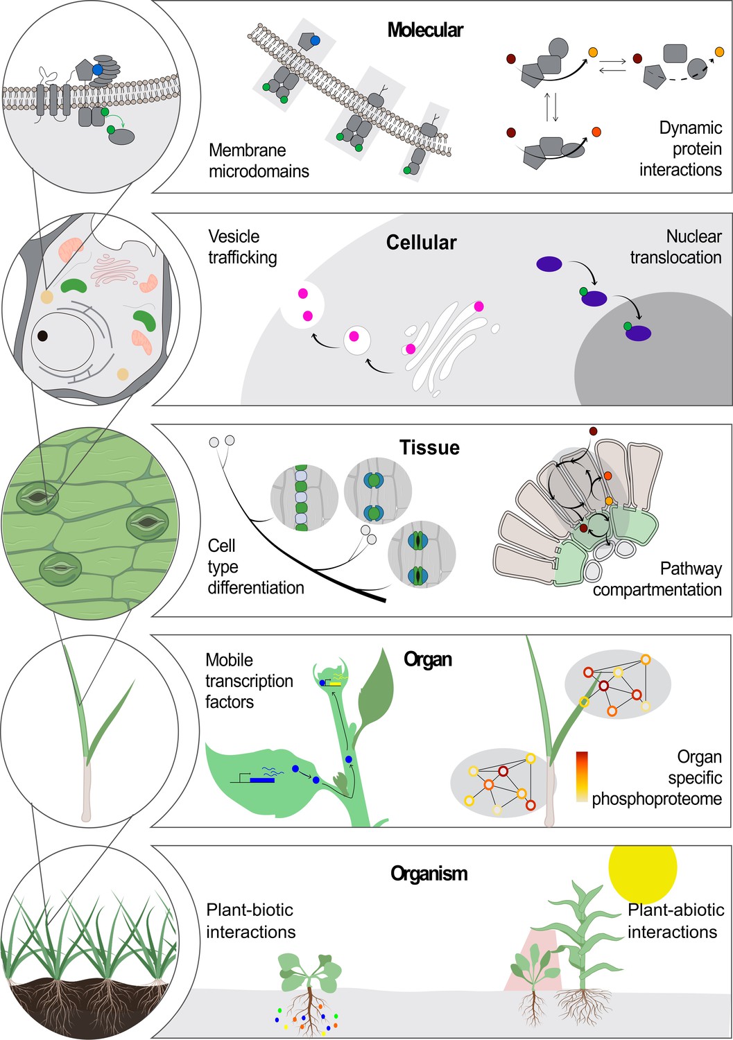
The relationship between the sequence, structure and function of a protein has been explored from many angles, from biophysics to drug discovery (Brausemann et al., 2017; Rajendran et al., 2018; Shang et al., 2020). This long-standing paradigm has driven the development of innovative technologies in imaging, mass spectrometry, genomics and bioinformatics (Camp et al., 2019; Mallis et al., 2020; Nakane et al., 2020). Yet an evolving paradigm stating that the location of molecules is essential for their function at all scales, from molecular complexes to cells, organs, organisms and whole communities, also holds true (Figure 1).
Figure 1
Location-to-function paradigm.
The illustration shows the levels of organization of a plant (left) and a few examples highlight how location determines function at each level (right). At the molecular level, functions of protein complexes can be determined by their localization in membrane microdomains (Jarsch et al., 2014) or by dynamic protein interactions (Obata, 2019). At the cellular level, a protein can be located differentially through transport mechanisms, including vesicle trafficking (Goring and Di Sansebastiano, 2018) and nuclear translocation (Marchive et al., 2013), regulating its function. At the tissue level, cell position can drive its fate into a specialized cell type (Shao and Dong, 2016). Metabolic pathways can operate specifically in specialized cell types across tissues (Marchive et al., 2013; Schlüter and Weber, 2020). At the next level, the existence of non-cell-autonomous transcription factors can transverse intercellular scales across plant organs (Han et al., 2014). Also, an organ-dependent post-translational proteome has been described as a mechanism of protein function regulation (Uhrig et al., 2019). At the organism level, plant interaction with biotic (Harrison et al., 2002) and abiotic factors (Michaud et al., 2017) can occur through a localized cue perception.
At the molecular level, compelling examples of the location-to-function paradigm include the multi-enzyme complexes that work as molecular machines (so-called ‘metabolons’) to channel intermediate metabolites and produce specific molecular products in a highly controlled manner (Obata, 2019).
At the cellular level, the partitioning of proteins and lipids into organelles – and even membrane microdomains within organelles – plays crucial roles in molecular trafficking and signal transduction, which have great impacts in many biological processes such as plant development and defence responses (Jarsch et al., 2014; Xing et al., 2019; Yu et al., 2020; Heinze et al., 2020; Shimizu et al., 2021). The differential location of a single molecule can determine one of several alternative functions (see, for example, Smirnoff and Arnaud, 2019). Protein function can also be dynamically regulated by a change in location, such as the activation of transcription factors by translocation into the nucleus (Jiang et al., 2020; Marchive et al., 2013; Ramon et al., 2019; Pastor-Cantizano et al., 2020). Furthermore, protein translocation to different cellular domains results in different functional outcomes (see for instance, Kim et al., 2020).
At the tissue level, C4 photosynthesis is a classic example of how differential molecular localization between cell types enhances plant performance: differential expression of enzymes in bundle sheath and mesophyll cells in C4 plants can concentrate CO2 in close proximity to Rubisco, which is not possible in C3 plants where the enzymes are present, but not differentially expressed between the cells (Schlüter and Weber, 2020).
Considering the opportunities in science and technology today, we contend that this ‘location-to-function’ paradigm will likely usher in conceptual leaps and transformative technologies, as was the case for the structure-to-function paradigm.
One particular area of potential success is agriculture, as the success of molecular approaches for crop improvement depends on a detailed understanding of the spatio-temporal regulation of genes and proteins and the production of metabolites (see Wang et al., 2008 for an example of how controlling the location of gene expression impacts rice grain size and yield). The timing of expression is also a key determinant in imparting functional specification and influencing traits. For instance, engineered early expression of WRINKLED1 (WRI1, an AP2 class transcription factor) during maturation increased Arabidopsis seed size and oil content (Kanai et al., 2016). More recently, an ambitious goal to globally sequester extra CO2 into roots was established, which predicates on understanding what controls the development of a specific tissue, periderm, and the cell-type-specific expression of suberin, a lipophilic polymer found in plant cell walls. These examples illustrate how a thorough understanding of the cell- or tissue-specific expression of plant genes may enable the development of more effective biotechnological tools to facilitate crop improvement, plant resilience and climate change mitigation. Currently, these spatially- or cell-type-resolved expression data are missing or incomplete for most crop plants and there is no centralized repository for their integration. These shortcomings limit the rapid development of improved crops based on location-to-function data.
Unlocking location-to-function: The first Plant Cell Atlas (PCA)
The PCA workshop
Establishing a comprehensive picture of where molecules are located and how their locations inform function in higher plants will require concerted efforts from diverse groups of scientists. To catalyze such a community, a series of workshops were organized by Carnegie Institution for Science, New York University and Stanford University scientists around the topic of a cell atlas for plants, a framework to generate and synthesize molecular and cellular data on the development, dynamic functions and specialization of plant cells. This effort would also strive to determine the state of the field, identify necessary components and people and discuss a path forward. Three workshops were held virtually during May and June of 2020. Over 400 scientists participated, of which 70% were early-career researchers, and 33% non-US based (Rice et al., 2020). Attendees of this first series of workshops became the basis of the PCA community. Following the success of the inaugural meeting, two technology-oriented workshops on single-cell sequencing and spatial proteomics and a career planning workshop were held virtually in 2021, respectively attracting 273, 253 and 42 participants. Additional workshops and symposia will be announced on the PCA website.
The PCA goals
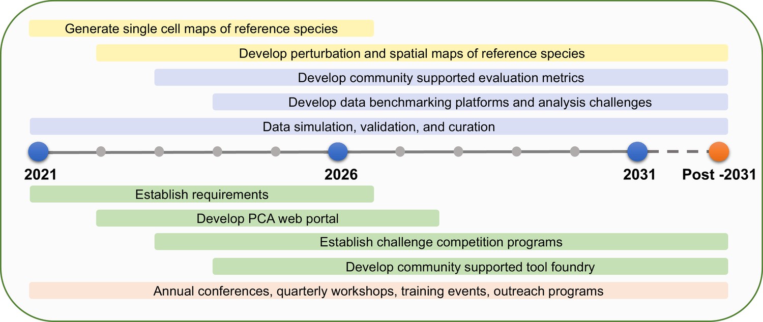
The goal of the PCA is to bridge gaps in knowledge, providing critical localization, dynamics and interaction information at the cellular and subcellular levels. The participants in the PCA Research Coordination Network envision a set of concrete milestones over the next decade to reach that vision, based on community goals set during these meetings in 2020 and 2021 (Figure 2).
Figure 2
PCA milestones.
PCA milestones for the next 10 years and beyond in data generation (yellow bars), data analysis (purple bars), software development (green bars), and building PCA community (orange bar).
Identify and quantify relative abundances of all definable cell types in every organ of several strategically selected reference species (e.g. a single-celled organism, a non-vascular plant, a eudicot, a monocot).
Profile the developmental trajectory of every specialized cell type in each organ in the reference species through single-cell sequencing.
Develop a ‘reference’ clearinghouse where new experiments based on single-cell profiling can be mapped onto comprehensive atlases representing the diversity of known transcriptional states among cell types.
Profile the responses of major organ systems to a common set of environmental perturbations at a single-cell resolution.
Determine the intraspecific variation of developmental trajectories and environmental responses at a single-cell resolution.
Establish data integration platforms and community-supported data standards to better enable data integration.
Develop a community-supported data benchmarking platform that will accomplish simulation and validation of data and evaluation metrics.
Determine protein subcellular localization, interactions and dynamics in the major cell types of leaves, roots and flowers for every gene in reference species.
Curate empirical datasets and develop data analysis and visualization tools through community-driven challenges (e.g., the DREAM single-cell transcriptomics challenge; Alonso et al., 2019; Pham et al., 2020).
Establish mechanisms for fostering new collaborations and funding for data generation, data integration as well as new technology and method development or adoption for plants.
Some of these goals can be accomplished quickly, as already existing technologies such as single-cell and single-nucleus RNA-seq make them realistic. Others require the development of technologies to collect information at the single-cell level. As technology and discovery reshape the vision of the PCA, new aspirations should emerge from the community. Nonetheless, these ambitious milestones represent key gaps in knowledge that will accelerate plant research (if addressed) and are feasible to achieve given the opportunities presented by recent technological advances. The PCA community can enable this vision in several ways.
The PCA vision
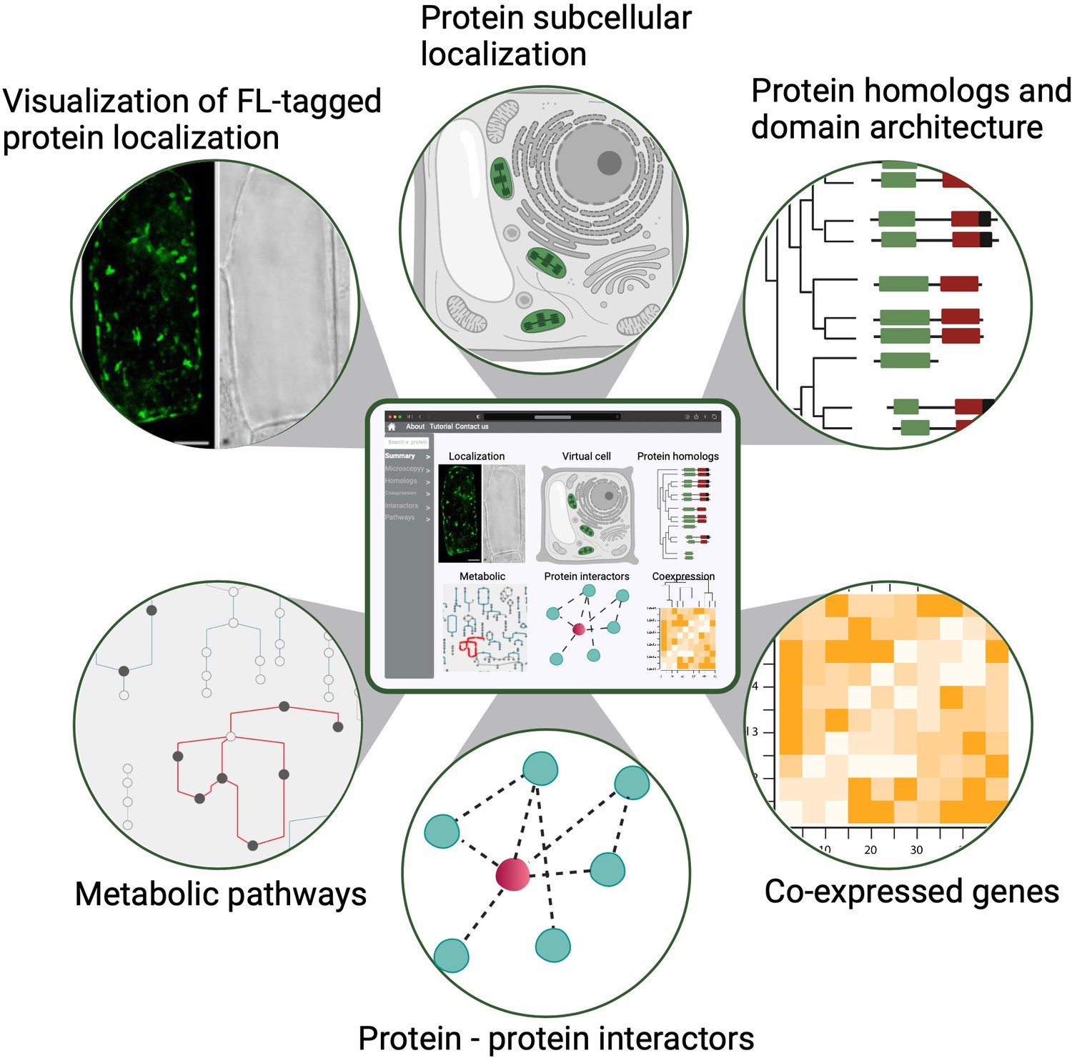
First, the participants in the PCA Research Coordination Network envision a one-stop, user-friendly website with illustrated and graphical details about cellular organization across different developmental stages. This information, modeled after findable, accessible, interoperable and reusable (FAIR) data principles, could be widely accessed and used to advance research and develop educational tools (Figure 3; Wilkinson et al., 2016). Although single-cell genomics data (mostly transcriptomic) have been generated and analyzed in individual studies for several years, how well the broader community can use these datasets in comparative analyses has been limited. This is largely due to the difficulty in integrating and comparing data generated by different methods, growth conditions and collection methods. The PCA will be critical in harnessing the power of single-cell data by establishing and promoting community-supported best practices in sample preparation, data generation, analysis and integration – including data benchmarking platforms, evaluation metrics and reference cell maps for major organ/cell types in model species. Important considerations for such infrastructure include how data will be gathered, curated and stored in order to integrate resources and contributions from different laboratories. It is necessary to develop and incentivize standards for both data collection (i.e. experimental protocols) and curation to allow integration of disparate datasets into a unified resource such as the PCA. This should include application programming interfaces (APIs) for programmatic access to ensure that PCA is accessible to all in a FAIR format. Similar standards have been produced for instance for plant phenotypic data (Papoutsoglou et al., 2020).
Figure 3
A conceptual diagram of a PCA user interface.
An example of a PCA user interface that integrates various data types from molecular, biochemical, cellular and evolutionary contexts to connect location to function. Shown are example data types that would be seamlessly connected to enable easy navigation and discovery (FL: fluorescence).
Second, we challenge and support the community to create a 4D representation of a developing plant from root to fruit (Henkhaus et al., 2020) with data collected from single-cell omics platforms (genomics, epigenomics, transcriptomics, proteomics and metabolomics) for each cell or cell type that are mapped onto tissue atlases of organ development. The data collected from the single-cell platforms form the basis of these atlases and serve as a reference for mapping further experimental data (e.g. from perturbation experiments). The PCA efforts include integration of not only single-cell omics data, but also spatiotemporal dynamics of single cells and their connection to cell types and developmental trajectories. It therefore facilitates a comprehensive understanding of how different complex tissues and organs are formed during plant growth and development.
Third, we wish to generate excitement and awareness about the potential for plant science research to address society’s most pressing challenges in agriculture, food security, bioenergy, resource management, as well as ecosystem stewardship and robustness in response to climate change. The PCA community needs to overcome the growing gap between ongoing research and societal perception with public communication and education. The PCA efforts should include outreach, training and education of the general public to understand the motivations, successes and limitations of this research. The initiative will help to build public awareness of how translational research that arises from model organisms can positively impact society and help feed the world. It will also demonstrate how understanding plants as foundational components of the ecosystem helps to predict and mitigate the consequences of climate change, and to be better stewards of the planet. Without these efforts, we fear that growing skepticism about science will impair the ability to aid society with the power of plant research, as apparent in the non-evidence-based regulation of gene editing in the European Union (Jorasch, 2020; Ruffell, 2018; Zimny et al., 2019).
Fourth, we see the PCA embracing new technologies and fostering multidisciplinary collaborations. The PCA will aim to build a community by introducing research tools to new users, and by facilitating interactions among scientists, engineers and breeders worldwide. These goals can be met by organizing in-person events and online workshops with presentations from domain experts, followed by breakout rooms for targeted discussions and networking. The multidisciplinary nature of the PCA has enormous potential for accelerating the fundamental understanding of plant cells and how they connect to the whole organism, and for translating fundamental discoveries to agriculturally important crops across the globe. The dream of the PCA initiative in this regard is to actively engage applied plant scientists and breeders by facilitating the formation of joint national and international funding schemes across scientific disciplines. This will allow diverse communities to benefit from each other's expertise and for ideas and technologies to be transferred from lab to land. Enhancement in research translatability will address globally relevant societal questions that are aligned with the UN Sustainable Development Goals (Biermann et al., 2017; Costanza et al., 2016; Griggs et al., 2013), including food sustainability (Goals # 1–3), resources to tackle climate change (Goal #13) and the overall survival of the biosphere (Goals # 14–15). We envision that starting with a comprehensive cell atlas of model organisms will help to better understand non-model species and eventually expand to an ecosystem-scale analysis of interactions between organisms.
Inspiration from similar projects
What the participants in the PCA Research Coordination Network envisage is ambitious and not yet available for any system. However, examples of components of our vision will serve as guides. Among the animal whole organism atlases (Lähnemann et al., 2020), the mouse brain and adult mouse cell atlases are models for comprehensive coverage and community accessibility (Rosenberg et al., 2018; Saunders et al., 2018; Zeisel et al., 2018; Tabula Muris Consortium, 2018; Han et al., 2018). The Human Cell Atlas’ data portal already includes data from 55 organs and 12 million cells, and it is becoming a model for making community-generated single-cell data available at a single portal, along with APIs and data analysis pipelines made easily accessible. In addition, the OpenWorm project provides an excellent example for modeling, visualizing and simulating various aspects of the biology and behavior of an organism (Szigeti et al., 2014). For plants, response to the environment would serve as an analogous level to behavior. EMBL-EBI’s Single Cell Expression Atlas recently started hosting plant single-cell transcriptome data, which is an exciting development (Papatheodorou et al., 2020). Finally, the Global Natural Product Social Molecular Networking initiative provides a model for a one-stop-shop web infrastructure to enable data generation, integration, analysis and publication from mass spectrometry (Wang et al., 2016).
The envisioned PCA community
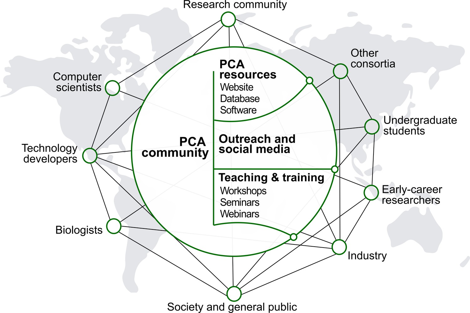
The PCA will be developed, maintained and used by a global community with diverse scientific, technical, creative and educational backgrounds (Figure 4). Helped by rapidly evolving online tools that increase inclusivity and transparency, the initiative should seek ways to facilitate the engagement, communication and collective decision-making among all groups involved. This work will aim to be grounded in active listening, psychological safety, teamwork, mutual respect and integrity. An inclusive and bottom-up ethos will underpin the development of global biological databases built with scientific rigor and transparency.
Figure 4
People and culture.
Major stakeholders of the PCA are described. The goal is to establish a broad network of collaboration between developers and users through the creation of an accessible platform, educational tools and outreach activities. The PCA community should strive to be inclusive, diverse and transparent.
The participants in the PCA Research Coordination Network anticipate that the success of the PCA will hinge on an open and frequent dialogue between developers and users, with facilitation and guidance from a steering group. Recruiting scientists from diverse disciplines as well as from different career stages will be crucial in the successful development of the PCA. In particular, the initiative should continue to provide an opportunity for early-career researchers and foster the development of future leaders in plant biology.
Developers
The mission of the PCA community will be achieved by groups who will contribute to the development and maintenance of the database, as well as those who generate or use the data and provide inputs for improvement. Developing the PCA database will be done with the expertise of scientists, software engineers, modelers, scientific illustrators and animators coming together to fine-tune the display of cellular organization and collecting information. Data and computational scientists will have to be involved in the curation and annotation of data. Engagement will extend to consortia pursuing similar activities in different biological systems (e.g., Human Cell Atlas). As the PCA will heavily depend on data visualization, user interface and experience design will also benefit from advisors in art and design fields. Additionally, collaboration with cartographers will enhance the understanding of spatial mapping for developing the PCA database. These influences from outside the field of plant research will complement and bolster the expertise of plant scientists in the PCA community.
Users
The participants in the PCA Research Coordination Network expect several types of users. One of the major user groups will be the data generators, which will involve plant scientists from many disciplines, including single-cell profiling, protein-protein interactions, cell and developmental biology, imaging or synthetic biology. To engage scientists from different career stages and areas of plant science, it will be important to develop mechanisms attributing and giving credit to data contributors. Another type of users will be researchers who wish to explore the PCA data to make discoveries. Engaging these users through a bottom-up process driven by their needs will be required for a vital and thriving PCA. Finally, the third group of users, both within and outside of the research community, are those who will access the PCA for educational purposes. Simple and intuitive interfaces combined with an attractive display of information and compelling visualizations will attract a large number and range of educators seeking information for teaching.
Integrating the PCA in the wider bioinformatics community
Robust computational infrastructure development will be essential to support the research, community-building and outreach activities of the PCA (Figure 5). We envision the PCA infrastructure (that is, the technologies, standards and computational platforms used by developers to build a framework to house and make available the PCA-relevant data) to have a seamless integration of multi-omics data with localization data at multiple scales of the plant. A governance structure, contribution guide and code of conduct for participants in infrastructure development should be established to define and support community values. These values include open-source development, FAIR principles and outcomes-based planning and implementation. Stakeholders should be included in the decision-making process. To implement these values, existing efforts with similar values and goals should be leveraged. For example, the DREAM challenges group could be leveraged for developing data analysis, visualization and access tools (Pham et al., 2020).
Figure 5
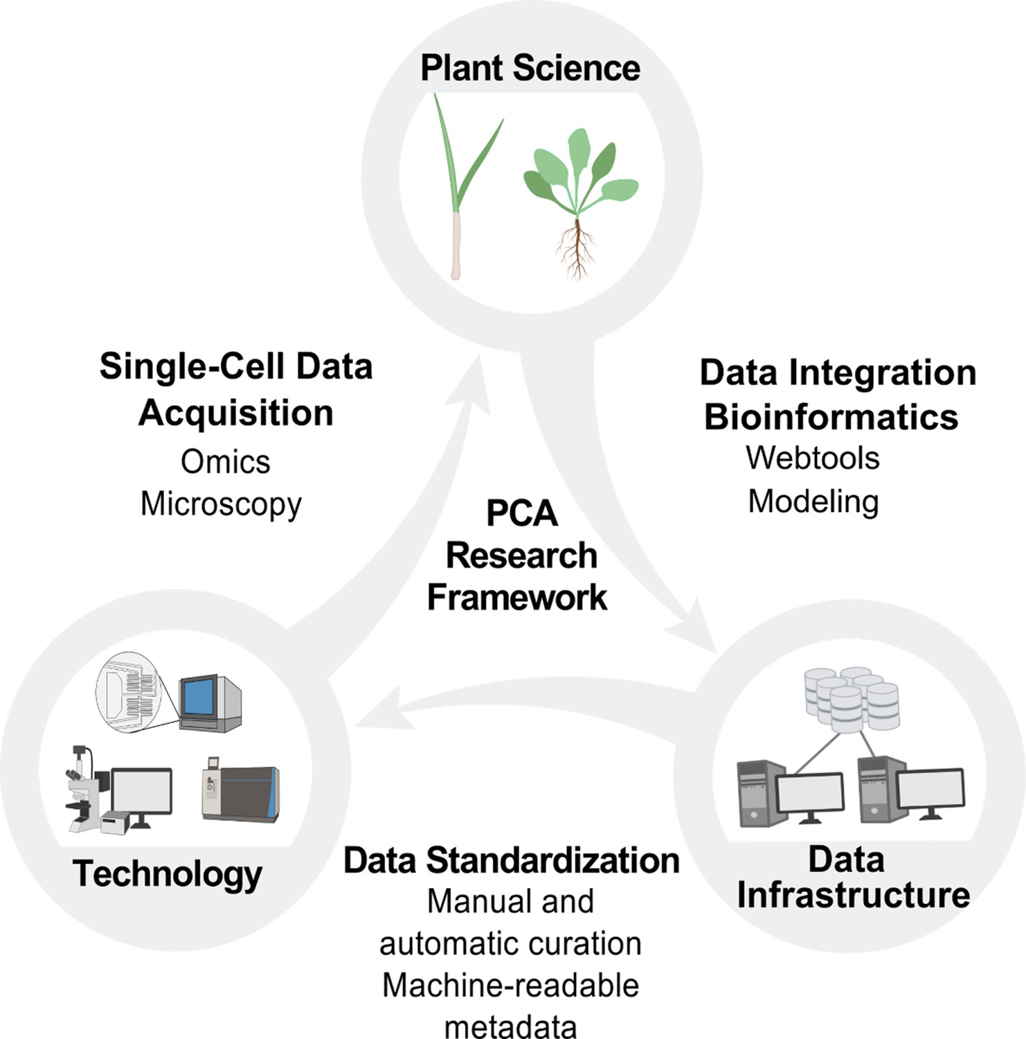
PCA research framework.
Building the PCA will require cooperation and coordination between plant science, technology and data infrastructure. Plant scientists will use new technologies to generate data at the single-cell level, which will be integrated by data scientists into the PCA infrastructure through manual and automated curation. This data integration will enable the development of new technologies and drive new hypotheses and investigations for plant scientists, who will then contribute additional data to the PCA resource.
While new tools and resources will be needed to build a PCA community platform, existing resources should be strong guiding principles when developing the PCA infrastructure. The PCA should make efforts to liaison with data repositories, knowledge bases and data analysis hubs such as ATTED, eFP Browser, ePlant, Ensembl Plants, Gramene, MaizeGDB, TAIR, The 1001 Arabidopsis Genomes phenotype database, NCBI’s GEO, EMBL-EBI’s Expression Atlas, AraPheno and others (Obayashi et al., 2007; Sullivan et al., 2019; Howe et al., 2020; Tello-Ruiz et al., 2021; Portwood, 2018; Berardini et al., 2015; Seren et al., 2016; Barrett et al., 2005; Papatheodorou et al., 2020; Waese et al., 2017). For example, OMERO is a global consortium effort aiming to standardize biological imaging data, which includes data depository as well as image processing software to translate image files into a universal format. Cross-species information may be mapped to the latest phylogenetic tree updated by the Tree of Life Web Project and Bioschemas. Engagement should also extend to consortia that are undertaking related work in other organisms, such as the Human Cell Atlas. To create a collaborative research environment, projects such as Synapse, Cyverse and KBase could be evaluated for strategic partnership. To develop a framework to represent the PCA at multiple scales, it will be important to implement conceptual frameworks such as the common coordinate framework (Rood et al., 2019), developmental trajectories (‘pseudo-time’) and spatial gradients (‘pseudo-space’; Stewart et al., 2019).
Driving the community forward
The PCA will receive diverse sets of data from different types of techniques and experiments, many of which are constantly pushing the boundaries of the state of the art. As such, the PCA needs to position itself and its community at the frontier of plant biology, looking out for emerging findings and technologies. This could be achieved through online workshops that enable pioneering researchers and developers to introduce emerging technologies to the community. Other training activities might include hands-on workshops on single-cell, spatially resolved experimental techniques, data annotation and analysis tutorials, as well as hackathons and virtual office hours to help novice code developers, data scientists and other researchers.
Gaps in knowledge: How the PCA could address unresolved questions in plant science
Many unresolved questions in plant biology require new technologies and approaches that the PCA could drive forward, as well as benefit from. Examples of such major knowledge gaps are described here (see also Figure 6).
Figure 6

Knowledge gaps to fill for the PCA.
Several limitations inherent to plants (shown as pieces of a jigsaw puzzle) must be overcome to enhance the understanding of plant cells as biological systems. To characterize the unique molecular attributes of each cell type composing a plant, plant biologists will need to develop reliable methods to broadly access the different cell types across various plant species. Also, single-cell multi-omics technologies will be necessary to get a deeper understanding of plant cell processes across all layers of molecular regulation. This data must be gained in the context of functional annotation of the cells. This would require the identification of reliable marker genes for each type of plant cell-. Ultimately, the spatiotemporal distribution of the molecular attributes of each cell will help to understand their dynamic regulation during plant cell development and in response to environmental stresses. Integrating the information collected using bioinformatics tools will enable the characterization of regulatory networks at a single-cell resolution and comparative analyses across plant species at the cellular level. This data will fuel the establishment of new synthetic biology strategies to enhance plant biology.
Plant development
The PCA could unlock answers to questions that require understanding the molecular changes in cells during development. The potential to characterize embryonic development comprehensively at single-cell resolution and in the multicellular context of complex tissues could help decipher the ‘rules’ underlying the earliest stages of plant tissue formation (ten Hove et al., 2015). In post-embryonic development, plant meristems are remarkable multicellular machines, continually producing new cells that must be specified to fate and then guided through phases of maturation (Esau, 1953; Sinnott, 1960). A major challenge is to understand the mechanisms that coordinate these cell maturation and fate specification events within meristems. Furthermore, plants present a fascinating contrast to mammalian systems with respect to the plasticity they maintain (Borges, 2008). For example, perennial plants often show meristematic plasticity, where only a subset of meristems per season are activated, and with the meristematic activity frequently modulated in response to external and internal cues (Tylewicz et al., 2018). The power of single-cell omics to capture the dynamic molecular coordination of tissue maturation at fine scales promises to help advance these classic questions in plant development (McFaline-Figueroa et al., 2020).
Response to the environment
As for problems in development, how plants respond to environmental stimuli requires understanding the molecular changes taking place in individual cells and how these changes are communicated cell to cell, across tissues and among organisms. An example can be found in how the threat of invasion of foreign biota into cellular and intercellular spaces often induces localized hypersensitive responses of programmed cell death (Jones and Dangl, 2006; Ngou et al., 2021). The question remains as to how the signals induced at the attack sites are communicated to neighboring cells and propagated to distal regions. Do hidden pre-patterned states among seemingly homogenous cells help the plant anticipate attacks? Or is there heterogeneity in cellular responses that could reveal a more complex response to infection? How do these processes differ between susceptible and resistant plant varieties? Heterogeneous responses to infection around invasion sites, as well as differences in molecular signals that correlate with cellular immunity, could provide new insights into how successful defense responses are coordinated.
A parallel set of questions emerge from biotic interactions involved in symbioses, such as the ancient association of land plants with mycorrhizal fungi, or more recent symbioses like those formed by members of the nitrogen-fixing Fabids clade (Wang and Qiu, 2006). In the latter case, infection by the symbiont through the root hairs takes place asynchronously, so bulk sampling often leads to profiles that are a heterogeneous mixture of cell states (Libault et al., 2010). Can new technologies help to gain a discrete view of how individual cells respond to the symbiont over time? Can fresh insights be gained into plant-symbiont interactions by studying root cells and specialized symbiont structures (i.e., arbuscules, nodules) simultaneously? In addition, can the chain of cellular communication in the plant throughout the stages of microbial colonization be better understood? Lastly, by comparing these processes across plants with differential symbiotic specificities, is it possible to start understanding the hallmarks of a robust symbiotic relationship?
Cellular evolution
From rhizoids to pollen, plants have evolved many new specialized cell types, allowing adaptation to new ecological niches. In addition, specific cell types in the root harbor molecular mechanisms to perceive and respond to environmental stimuli, suggesting they may act as ‘gatekeepers’ for these responses (Henderson and Gilliham, 2015). What new perspectives on cellular evolution could we gain by producing comparable single-cell omics data sets on strategically selected species within the plant kingdom? Previous attempts to use expression patterns of homologous genes in distantly related species to assess functional orthology have provided insights (Adamski et al., 2020). However, bulk tissues collected in different species may have substantially different composition of cell types (Huang and Schiefelbein, 2015). Furthermore, the presence of novel cell types in some species may be obscured in whole organ samples (Kajala et al., 2021). By focusing on the fundamental unit of organs – cell types and cell states – comparative studies of species representing key plant lineages could identify minimal molecular toolkits driving conserved plant cellular functions. In addition, these single-cell approaches could highlight the differences in developmental pathways that lead to phenotypic diversity and adaptation to a myriad of environmental conditions.
Technical challenges faced in the development of the PCA
Critical challenges must be overcome to achieve the data generation, analysis and software development milestones of the PCA (Figure 2). Many mirror those faced by the larger single-cell initiatives that include animal models (Lähnemann et al., 2020), but we focus here on those that specifically affect the plant research community (summarized in Box 1).
Box 1.
Major challenges in building a PCA.
Isolating plant cells and subcellular compartments. Efficient strategies for dissociating plant cells away from their structural context are needed to understand biological processes at the single-cell level. The fidelity and utility of methods requires extensive validation for collecting single-cell information across tissues and species.
Lack of well-characterized, cell-type-specific marker genes in non-model species. The functional characterization of plant cell types based on their transcriptomes suffers from the small pool of plant marker genes. The major limitation of the most widely applicable technology (Visium, 10x Genomics) is its relatively coarse resolution that would obscure the boundaries between several cell layers in plant tissues.
Incomplete annotation of plant genomes. Approaches that account for gene duplication, gene loss, and the potential for functional swapping among paralogs are required in plants. Comparison of individual transcript isoforms is difficult with current technologies. Efforts are needed to develop long-read technology to single-cell applications, or other strategies that can provide sequencing coverage to whole transcripts.
Need to integrate multiple components of the cell, such as nucleic acids (DNAs and RNAs), proteins, lipids, and metabolites. Addressing this challenge requires the development of multi-omics capabilities at cellular resolution, coupled with computational approaches that can integrate the data (Libault et al., 2017).
Plasticity of plant phenotypes. Plant phenotypes can be highly plastic according to changes in the environment. Efforts to establish reference atlases should ensure that standards in metadata description for growth conditions are established and adhered to.
Need to develop innovative strategies to functionally characterize plant genes at the single-cell level. Single-cell genetic technologies should be adapted to plant cells for high-throughput characterization of gene function in specific cells and cell types. For example, combining CRISPR-Cas9 technology with cell-type-specific inducible systems enabled the creation of genetic mosaics (Decaestecker et al., 2019; Wang et al., 2020). In addition, studies of proteins are limited to the whole plant-based tissue analyses. Approaches are needed for investigating single-cell or cell-type-specific proteomics in a high-throughput manner (Balasubramanian et al., 2021).
Limited resources. The plant science community is small compared to its animal or human counterparts and having access to more resources is a long-term challenge. Despite this, this community is relatively open and collaborative, making efforts to standardize aspects of single-cell experimental and analytical strategies potentially an easier barrier to surmount.
Isolating individual plant cells
A major challenge is dissociating plant cells: due to their rigid cell walls, some tissues and many species are recalcitrant to digestion (Himmel et al., 2007; Yoo et al., 2007). This feature limits the ability to sample mature tissues that often mediate environmental stresses. In addition, it can be difficult to isolate even young, meristematic cells in some key species that present cellular innovations. One emerging approach that has the potential to address this issue in plants is single-nucleus profiling, which avoids the need for cell wall digestion (Giacomello, 2021). Such nuclear isolation methods have already shown promise (Farmer et al., 2021; Marand et al., 2021). Recently developed proximity labeling approaches bypass the need for isolating cells or organelles by tagging proteins and RNAs near a target protein in living cells and organisms. These methods can profile components of protein complexes, dynamic protein-protein interactions, specific cell types and subcellular locations of proteins or RNAs (Zhang et al., 2019a; Mair et al., 2019; Huang et al., 2020; Kim et al., 2019; Wang et al., 2019).
Tracking cell provenance
Another challenge is mapping cells (or nuclei) back to their tissue location in the whole plant. Methods that can rapidly and conveniently generate ‘ground truth’ markers of cell types in situ will be especially important as more tissues and species with few existing markers are profiled. Adapting so-called spatial transcriptomic approaches to plants will be a critical tool. In particular, untargeted methods (that is, methods based on unbiased sequencing rather than hybridization of a limited set of predefined probes) have the potential for the resolution and throughput needed (Giacomello et al., 2017). In addition, nomenclature systems will be necessary to link the reference maps from different species that have varying organ anatomy and specialized cell types. The hierarchical approach proposed by the common coordinate framework for humans can serve as a starting point (Rood et al., 2019). However, unlike the coordinate systems used in animal organ atlases which focus on accounting for differences between individuals, plant coordinate systems should instead be geared toward discovering differences among representative species and populations. Any such system should be able to accommodate differences in cell type composition (Huang and Schiefelbein, 2015) and the development of novel cell types (Kajala et al., 2021).
Gene homology
Another set of issues surrounds gene homology in comparisons across populations and species. Plants have a well-known capacity for duplication at many scales, ranging from single gene to whole genome. An informative molecular profiling of cells will require high-quality genome annotations that consider the gene family expansion, gene loss and complex orthology relationships which frequently occur when extensive gene duplication follows speciation. Subfunctionalization and neofunctionalization can complicate assumptions about functional orthologs. On the other hand, difficulty identifying distant homologs that share low sequence similarity could obscure functional conservation. Advancement in sequence alignment methods, such as incorporating structural information, could help to discover these distant homologs (Morton et al., 2020). This will probably be an iterative process, where increased resolution of gene expression at the highly resolved cellular or subcellular level could allow inferences about functional orthology across species in large gene families.
Building the database and infrastructure of the PCA
Collection, standardization, curation, integration and visualization of data are key elements of a PCA platform, and the core PCA infrastructure should function as a data resource onto which services and tools are built.
Experimental and analytical standards
Establishing a vetted and community-approved set of experimental and analytical standards will be important. Such an approach was key to the success of the Encyclopedia of DNA Elements project, which ensured that data from multiple international laboratories were easily compared and integrated (ENCODE Project Consortium, 2011). Likewise, the 1001 Arabidopsis Genomes website provided a user-friendly interface to download all sequencing data as phases were completed – allowing the community to begin analyses and discoveries even before publication – and was complemented with an interactive tool to explore phenotypes (Seren et al., 2016) and their genomic associations (Togninalli et al., 2018). The standards were made easily available and updated over time as technologies advanced.
These platforms serve as data infrastructure blueprints as well as knowledge banks and inspiration. This would allow the PCA to be extended from an atlas of a single reference species and clone, to a set of atlases capturing natural variation (within and between species) in cell types and functions. For example, the interactive PCA platform could hyperlink the gene found in each tissue or cell type to the web portal of that gene in natural variation panels of the species of interest (such as the AraGWAS Catalog, Togninalli et al., 2020). Data structure scaffolds in the PCA could also be created so that the template can be replicated across genotypes of a species (e.g. different ecotypes) or different species, and allow users to select their desired organism or compare between the atlases.
Data visualization and exploration tools
In addition to data analysis and interpretation, the PCA infrastructure should include tools that provide effective ways to visualize real or virtual plant cells, and the dynamic processes that form and define them. This could enable hypothesis-generating in silico experiments that predict how plant cell types would respond to stimuli, or the properties of cells with a set of desired features. Creating these digital cells, tissues and other structures would require developing modeling, simulation, artificial intelligence, as well as data visualization and exploration tools similar to those being set up for other projects such as the OpenWorm (Sarma et al., 2018). Computational modeling and simulation-based predictions could then inform experimental work, the results of which could feed back into the PCA to improve models and predictions (Radivojević et al., 2020; Zhang et al., 2020).
Curating the PCA databases
To achieve the PCA’s vision of building a 4D representation of a developing plant, a wealth of existing data, resources and tools will need to be leveraged. For example, it is now routine to deposit next-generation sequencing, processed expression data, proteomics, metabolomics and protein structures in numerous repositories worldwide (Deutsch et al., 2017; Kaminuma et al., 2010; Leinonen et al., 2011; NCBI Resource Coordinators, 2018; wwPDB consortium, 2019). A combination of automated searches and manual curation of datasets from existing data repositories can be used to build the PCA data infrastructure. Reference genome databases such as TAIR, Phytozome, Gramene, EnsemblPlants and others that provide gene and pathway annotation resources (e.g. Gene Ontology) could enable an initial assessment of gene structure, location and function (Berardini et al., 2015; Goodstein et al., 2012; Howe et al., 2020; Tello-Ruiz et al., 2021; The Gene Ontology Consortium, 2019).
Building a platform that brings these data together with data exploration and visualization tools that allow a user to explore at multiple scales, from molecular to cellular to organismal, is a challenge. To this end, single-cell datasets should be made available in both raw and processed data formats and linked from the public-facing PCA website. This resource could additionally be populated using automated literature search tools, followed by manual curation by the PCA community. A selection of high-quality ‘reference’ single-cell datasets directly hosted by the PCA may also prove to be useful for benchmarking purposes. For all datasets, curation standards should be imposed to include minimal metadata falling into several categories: (1) experimental metadata should include a project name, details regarding how plants were grown, treated and sampled, as well as quality control metrics and definitions used to evaluate sample integrity; (2) cell metadata should include a cell identifier (where applicable), cell type annotations and other cell-level data that have come from standard or specific analysis of the dataset; and (3) gene metadata that include structural and functional annotations should be required.
In addition to providing links to data repositories, the PCA portal should include web-based tools to generate, access and query relevant data and metadata using a standardized ontology for metadata, similar to the Single Cell Expression Atlas (Papatheodorou et al., 2020) and the Human Cell Atlas implementation of metadata types. This will facilitate interoperability of datasets. Depending on the data type, scripts that implement standardized pipelines for single-cell data analysis could also be made available that reflect the current best practices, including those that implement methods for cross-platform and multimodal data integration as well as label transfer (e.g., Lotfollahi et al., 2020; Hie et al., 2019; Korsunsky et al., 2019; Kang et al., 2021; Stuart et al., 2019). Version-controlled source code that demonstrates analyses using the information in the PCA as a starting point should also be made available. These resources should promote greater use of the rapidly growing number of single-cell datasets.
Multiscale networks can provide an intuitive method to discretize and integrate different data types (Duran-Nebreda and Bassel, 2017). Other informatics frontiers that will be useful to explore include multi-dimensional image visualization and analysis tools such as Napari and Squidpy (Sofroniew et al., 2021; Palla et al., 2021), graph-based knowledgebases for integrating datasets (e.g., Reactome; Fabregat et al., 2018), natural language processing for automating curation (Braun and Lawrence-Dill, 2020), as well as machine learning and artificial intelligence methods for integrating heterogeneous data (Ma et al., 2020).
Challenges to the PCA infrastructure
Despite the widespread availability of data types described above, additional data that need to be integrated to achieve the PCA vision (metabolomics, imaging and phenotypic observations) are currently available in a variety of heterogeneous sources such as data sharing platforms (e.g. EMBL-EBI, Dryad, Figshare, and Zenodo), institutional data repositories, researcher websites or embedded within scientific publication records. A lack of centrality in data deposition makes these data less accessible.
In addition, methods for data standardization and curation will be critical tools to mitigate challenges with existing data, create best practices for collecting new data and ensure consistency to allow for data exchange and interoperability (Sansone et al., 2012). Experiments, datasets and other resources should be described in sufficient detail to be easily accessible and reproducible. Validating data with resource description frameworks is a means to ensure data compliance against the conceptual model it follows, data consistency and completeness. Data records and metadata should be machine-readable by using standardized vocabularies (i.e. cell type ontologies; Bard et al., 2005) to enable interoperability. Existing solutions and concepts of such ontologies are broadly applied in vertebrates and are available for plants, though they need to be widely adopted by the PCA community (Avraham et al., 2008; Bard et al., 2005 ; Diehl et al., 2016; Ilic et al., 2007; Xia and Yanai, 2019). Mapping ontologies to the 4D representation of the PCA will facilitate linking and visualizing the growing knowledge of plant cells.
Originality of the PCA infrastructure
The long-term vision for the PCA infrastructure is similar in scope to other moonshot ideas such as the Human Cell Atlas (Regev et al., 2017), the Transparent Plant (Henkhaus et al., 2020) and the Virtual Plant (Kost, 2001). However, the PCA vision is different in several important ways. The Human Cell Atlas is for a single organism with over 200 cell types, whereas the PCA community embraces cell atlas data from a diverse array of plants. A typical plant has about 100 cell types (https://www.ebi.ac.uk/ols/ontologies/po; Ilic et al., 2007). Understanding the diversity and origin of cell types across the plant lineage will be a central part of the initiative, as illustrated in Box 2 with the chloroplast as an example. The PCA is an essential step towards realizing the vision of a Transparent Plant, a simulated environment where virtual plants can be built from parts and behaviors (Henkhaus et al., 2020). In order to materialize such a simulation environment, where the parts are and how they work together must be understood. Finally, we see the Virtual Plant database (Katari et al., 2010) as an essential component of the PCA vision and infrastructure.
Box 2.
The chloroplast structure, function and dynamics illustrate PCA needs.
As highlighted here, technical solutions are needed to overcome key questions and challenges in chloroplast biology. The chloroplast is an important organelle in the plant kingdom, which is responsible for harvesting and converting light into chemical energy and sequestering carbon dioxide. Whereas much is known about chloroplast form and function, many aspects of chloroplast biology remain to be elucidated, especially in the context of the ‘location-to-function’ paradigm and proteostasis (Figure 7). The chloroplast is organized into several compartments (Solymosi et al., 2018; Figure 7A). This suborganellar compartmentalization allows for highly efficient and specialized metabolism, optimized light-driven electron transport, redox reactions and more (Rolland et al., 2018). The function and organization of chloroplasts greatly varies across the plant lineage and cell types (Pinard and Mizrachi, 2018; Figure 7B). For example, microalgae typically have a single chloroplast with a carbon-concentrating organelle called the pyrenoid (Barrett et al., 2021). In plants that can perform C4 photosynthesis, the chloroplasts in the bundle sheath and mesophyll cells have different compositions and functions (Edwards et al., 2004). A few plants even perform C4 photosynthesis within individual chlorenchyma cells where C4 function is obtained by spatial separation of function between dimorphic chloroplasts located in central and peripheral cytoplasmic compartments within the same cells (Erlinghaeuser et al., 2016; Edwards et al., 2004). In most land plants, the epidermal cells of leaves (except in guard cells) are devoid of chloroplasts but instead contain non-photosynthetic plastids, whereas aquatic plants are enriched in chloroplasts in their epidermal cell layer (Larkum et al., 2017; Han et al., 2020; Figure 7C). Recently Arabidopsis epidermal plastids have been shown to act in defense equipped with plant immune components (Irieda and Takano, 2021). Furthermore, different environmental conditions change the chloroplast proteome as well as relative positions of chloroplasts and their parts at various scales (Kong and Wada, 2014; Figure 7D). Finally, the chloroplast life cycle includes various transformations in form and function from birth to differentiation to senescence, and how these transformations occur is an understudied frontier (Llorente et al., 2020; Solymosi et al., 2018; Figure 7E). It is still not fully understood how chloroplast and other types of plastids differentiate, adapt, function and integrate their activities within specific cell types. Single-cell technologies bring promise for solving key challenges in cell-type and species-specific differentiation of plastids. Examples of key technologies and how they could contribute to a range of PCA-related question are listed below:
Affinity tagging of plastids using cell-type-specific promotors driving the expression of tagged proteins that associate to the plastid surface. Recently this approach has been developed for Arabidopsis thaliana mitochondria (Niehaus et al., 2020; Kuhnert et al., 2020). Successful cell-type-specific plastid affinity purification would resolve molecular features, properties and associated functions of plastid with high precision. This could include determination of plastid RNA, protein and metabolite populations through high sensitivity, state of the art RNA-seq profiling and mass spectrometry techniques.
Time-resolved plastid-targeted proximity labeling techniques to understand intra-plastid protein organization and dynamics. Recently, proximity labeling approaches have been successfully implemented in plants. They enable discovery of proteins that interact with specific regions of a bait protein, as well as identify localized protein interactions at high spatial and time-resolved resolutions (Huang et al., 2020; Mair et al., 2019; Zhang et al., 2019b). This novel approach would enable resolving intra-plastid protein organization and dynamics for different cell types and photosynthetic and non-photosynthetic plastids.
Technology to detect and track retrograde signaling pathways from plastids to nuclei in response to, for example, singlet O2, protein folding stress, redox and metabolites. These retrograde pathways involve a range of small molecules and likely also protein signaling cascades (Jiang and Dehesh, 2021; Muñoz and Munné-Bosch, 2020; Pesaresi and Kim, 2019). Non-invasive, high-resolution and high-sensitivity imaging technologies should be developed or adapted to address this challenge.
Plastid-localized reporter systems (biosensors) to monitor pH, calcium, ATP and redox potential. There are now many such reporters (Isoda et al., 2021; Okumoto and Versaw, 2017) and a fluorescent ATP sensor protein that allows the measure of ATP concentrations in the plastid and cytosol (Voon et al., 2018). Functional relationships between biophysical and biochemical parameters could be probed through the systematic application of reporter systems in plastids.
Plastid protein lifetime reporter technologies to understand cell-type-specific proteostasis. A novel protein lifetime reporter system has only been applied to address N-degron pathways in the cytosol (Zhang et al., 2019b). Adaption to plastids would resolve the complexities of plastid proteome remodeling in response to development and (a)biotic changes (Bouchnak and van Wijk, 2019).
Synthetic biology to engineer novel plastid metabolic pathways. Plastids are excellent targets for metabolic engineering through synthetic biology: they have their own genome and generally tolerate high levels of ‘foreign’ protein without significantly impacting function or proteostasis (Boehm and Bock, 2019; Jensen and Scharff, 2019).
Figure 7
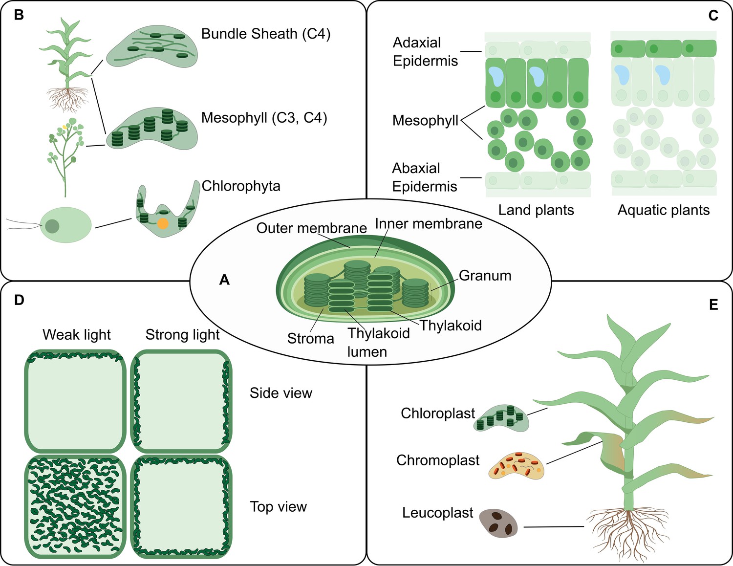
The chloroplast structure and function in connection to location.
The PCA could facilitate an increased understanding of the biology of the chloroplast by providing accessible, integrative and spatially-resolved data for transcripts, proteins and metabolites across cell types (Box 2). (A) A representation of a typical chloroplast structure and its compartments. (B) The chloroplast structural differences in some plant lineage. (C) A leaf cross-section showing different chloroplast location at the tissue level in land and aquatic plants. (D) The chloroplast location can change within the cell upon environmental cues such as light intensity. (E) Various forms of chloroplasts in different organs.
Potential sources of funding for the PCA
The PCA will require adequate funding to achieve its full potential – including translational impact for agriculture (Bartuska, 2017; Bol et al., 2018; Gök et al., 2016; Hu, 2020; Jaffe et al., 2015; Rosenbloom et al., 2015). As the PCA has an international reach, programs that facilitate collaboration across borders will be valuable in advancing the goals of the initiative, such as partnering programs by the Biotechnology and Biological Sciences Research Council (BBSRC) in the United Kingdom, European Commission’s Horizon Europe and the United States’ Fulbright scholars program. Similar programs exist that foster alliances between scientists from the US with those in Japan, Israel and Germany; and between India and other countries. As big data, nanotechnology, computation and artificial intelligence have now become an integral part of biological research, funds to support the interdisciplinary vision of PCA will be important to foster integration of these disciplines.
While governments are the major sources of funding for basic research, their level of funding has been steadily decreasing (Khan et al., 2020). Foundations are the major source of private basic research funding and are less constrained by time, politics and topic. They have a high potential for funding initiatives in emerging areas such as the PCA. However, it is challenging for foundations and individuals to find worthy initiatives. To fill this gap, the Science Philanthropy Alliance was established. One of their success stories is the Chan Zuckerberg Initiative providing substantial funds to develop technologies for the Human Cell Atlas initiative. Such a partnership for the PCA would take its vision to new heights.
Industry is another potential source of funding. Over 70% of applied research and development in the US is funded by industry (Khan et al., 2020). A thorough understanding of plant systems is essential for designing effective agro-biotech solutions leading to new crop varieties, and to innovative crop protection products that can leverage sustainable food production. A partnership with the PCA initiative would accelerate development of new solutions for customers of the agbiotech industry. Thus, funding and collaborations, ranging over multiple disciplines and countries, and multiway interactions amongst academia, industry and philanthropy will be essential to realize the vision of the PCA.
Conclusion and outlook
Future economies will be increasingly plant-based. Biomass is projected to be a major source of primary energy by 2050 (Reid et al., 2020), plant-based meat and dairy products are already transforming the food industry (Shepon et al., 2018), and plant-based vaccines against several diseases are increasingly being researched (reviewed by Shahid and Daniell, 2016). With the rapidly changing climate, economy and values in our society, now is the time to reimagine plant science as a critical component of the future not only for agriculture, but also the environment, energy, health care, manufacturing and technology.
Today, plant science constitutes a minority of life sciences in terms of funding and workforce. To meet the demands of the future economy, we need to strengthen plant science, attract talent from other fields and train the next generation of plant scientists (Henkhaus et al., 2020). In 2004, the evolutionary biologist John Avise wrote that ‘‘despite nearly three decades of experience with recombinant DNA techniques, the ultimate contribution to the broader human enterprise remains uncertain’’ (Avise, 2004). Two decades after this statement, in 2020, recombinant DNA technology ushered in the beginning of an end to a global pandemic that cost millions of lives, through a record-breaking runtime of development and deployment of mRNA and DNA-based viral vaccines. While still at its infancy, we hope that the PCA initiative has the potential to serve as a nucleator, bringing together scientists and engineers from a wide range of fields to solve fundamental problems in plant biology with innovative solutions from emerging technologies.
Data availability
No associated datasets.
References
-
NCBI GEO: mining millions of expression profiles-database and toolsNucleic Acids Research 33:D562–D566.https://doi.org/10.1093/nar/gki022
-
Pyrenoids: CO2-fixing phase separated liquid organellesBiochimica Et Biophysica Acta (BBA) - Molecular Cell Research 1868:118949.https://doi.org/10.1016/j.bbamcr.2021.118949
-
Global governance by goal-setting: the novel approach of the UN sustainable development goalsCurrent Opinion in Environmental Sustainability 26-27:26–31.https://doi.org/10.1016/j.cosust.2017.01.010
-
Plasticity comparisons between plants and animals: concepts and mechanismsPlant Signaling & Behavior 3:367–375.https://doi.org/10.4161/psb.3.6.5823
-
N-Degron pathways in plastidsTrends in Plant Science 24:917–926.https://doi.org/10.1016/j.tplants.2019.06.013
-
The UN sustainable development goals and the dynamics of well-beingFrontiers in Ecology and the Environment 14:59.https://doi.org/10.1002/fee.1231
-
The ProteomeXchange consortium in 2017: supporting the cultural change in proteomics public data depositionNucleic Acids Research 45:D1100–D1106.https://doi.org/10.1093/nar/gkw936
-
The cell ontology 2016: enhanced content, modularization, and ontology interoperabilityJournal of Biomedical Semantics 7:44.https://doi.org/10.1186/s13326-016-0088-7
-
Bridging scales in plant biology using network scienceTrends in Plant Science 22:1001–1003.https://doi.org/10.1016/j.tplants.2017.09.017
-
Single-cell C(4) photosynthesis versus the dual-cell (Kranz) paradigmAnnual Review of Plant Biology 55:173–196.https://doi.org/10.1146/annurev.arplant.55.031903.141725
-
Reactome graph database: efficient access to complex pathway dataPLOS Computational Biology 14:e1005968.https://doi.org/10.1371/journal.pcbi.1005968
-
A new era for plant science: spatial single-cell transcriptomicsCurrent Opinion in Plant Biology 60:102041.https://doi.org/10.1016/j.pbi.2021.102041
-
The impact of research funding on scientific outputs: evidence from six smaller european countriesJournal of the Association for Information Science and Technology 67:715–730.https://doi.org/10.1002/asi.23406
-
Phytozome: a comparative platform for green plant genomicsNucleic Acids Research 40:D1178–D1186.https://doi.org/10.1093/nar/gkr944
-
Protein and membrane trafficking routes in plants: conventional or unconventional?Journal of Experimental Botany 69:1–5.https://doi.org/10.1093/jxb/erx435
-
Transcription factor-mediated cell-to-cell signalling in plantsJournal of Experimental Botany 65:1737–1749.https://doi.org/10.1093/jxb/ert422
-
BookThe “gatekeeper” concept: cell-type specific molecular mechanisms of plant adaptation to abiotic stressIn: Laitinen R. A. E, editors. Molecular Mechanisms in Plant Adaptation. John Wiley & Sons, Inc. pp. 83–115.https://doi.org/10.1002/9781118860526.ch4
-
Efficient integration of heterogeneous single-cell transcriptomes using scanoramaNature Biotechnology 37:685–691.https://doi.org/10.1038/s41587-019-0113-3
-
Ensembl genomes 2020-enabling non-vertebrate genomic researchNucleic Acids Research 48:D689–D695.https://doi.org/10.1093/nar/gkz890
-
The effect of public funding on research outputThe New Zealand Marsden Fund 52:1–22.https://doi.org/10.1080/00779954.2017.1325921
-
Engineering of plastids to optimize the production of high-value metabolites and proteinsCurrent Opinion in Biotechnology 59:8–15.https://doi.org/10.1016/j.copbio.2019.01.009
-
DDBJ launches a new archive database with analytical tools for next-generation sequence dataNucleic Acids Research 38:D33–D38.https://doi.org/10.1093/nar/gkp847
-
Extension of oil biosynthesis during the mid-phase of seed development enhances oil content in Arabidopsis seedsPlant Biotechnology Journal 14:1241–1250.https://doi.org/10.1111/pbi.12489
-
VirtualPlant: a software platform to support systems biology researchPlant Physiology 152:500–515.https://doi.org/10.1104/pp.109.147025
-
Recent advances in understanding the molecular mechanism of chloroplast photorelocation movementBiochimica Et Biophysica Acta (BBA) - Bioenergetics 1837:522–530.https://doi.org/10.1016/j.bbabio.2013.12.004
-
Towards a virtual Arabidopsis plantGenome Biology 2:REPORTS4019.https://doi.org/10.1186/gb-2001-2-8-reports4019
-
Rapid single-step affinity purification of HA-tagged plant mitochondriaPlant Physiology 182:692–706.https://doi.org/10.1104/pp.19.00732
-
Carbon-concentrating mechanisms in seagrassesJournal of Experimental Botany 68:3773–3784.https://doi.org/10.1093/jxb/erx206
-
The European Nucleotide ArchiveNucleic Acids Research 39:D28–D31.https://doi.org/10.1093/nar/gkq967
-
Root hair systems biologyTrends in Plant Science 15:641–650.https://doi.org/10.1016/j.tplants.2010.08.010
-
Plant systems biology at the single-cell levelTrends in Plant Science 22:949–960.https://doi.org/10.1016/j.tplants.2017.08.006
-
Integrative methods and practical challenges for single-cell multi-omicsTrends in Biotechnology 38:1007–1022.https://doi.org/10.1016/j.tibtech.2020.02.013
-
Development of native MS capabilities on an extended mass range Q-TOF MSInternational Journal of Mass Spectrometry 458:116451.https://doi.org/10.1016/j.ijms.2020.116451
-
The promise of single-cell genomics in plantsCurrent Opinion in Plant Biology 54:114–121.https://doi.org/10.1016/j.pbi.2020.04.002
-
Oxylipins in plastidial retrograde signalingRedox Biology 37:101717.https://doi.org/10.1016/j.redox.2020.101717
-
Database resources of the national center for biotechnology informationNucleic Acids Research 46:D8–D13.https://doi.org/10.1093/nar/gkx1095
-
Metabolons in plant primary and secondary metabolismPhytochemistry Reviews 18:1483–1507.https://doi.org/10.1007/s11101-019-09619-x
-
Expression atlas update: from tissues to single cellsNucleic Acids Research 48:D77–D83.https://doi.org/10.1093/nar/gkz947
-
Enabling reusability of plant phenomic datasets with MIAPPE 1.1New Phytologist 227:260–273.https://doi.org/10.1111/nph.16544
-
Functional diversification of ER stress responses in ArabidopsisTrends in Biochemical Sciences 45:123–136.https://doi.org/10.1016/j.tibs.2019.10.008
-
Unsung and understudied: plastids involved in secondary growthCurrent Opinion in Plant Biology 42:30–36.https://doi.org/10.1016/j.pbi.2018.01.011
-
The maize multi-genome genetics and genomics databaseNucleic Acids Research 1:D1146–D1154.https://doi.org/10.1093/nar/gky1046
-
A machine learning automated recommendation tool for synthetic biologyNature Communications 11:4879.https://doi.org/10.1038/s41467-020-18008-4
-
Aminoacyl-tRNA synthetases: structure, function, and drug discoveryInternational Journal of Biological Macromolecules 111:400–414.https://doi.org/10.1016/j.ijbiomac.2017.12.157
-
The main functions of plastidsMethods in Molecular Biology 1829:73–85.https://doi.org/10.1007/978-1-4939-8654-5_5
-
OpenWorm: overview and recent advances in integrative biological simulation of Caenorhabditis elegansPhilosophical Transactions of the Royal Society B: Biological Sciences 373:20170382.https://doi.org/10.1098/rstb.2017.0382
-
Regulation and evolution of C4 photosynthesisAnnual Review of Plant Biology 71:183–215.https://doi.org/10.1146/annurev-arplant-042916-040915
-
AraPheno: a public database for Arabidopsis thaliana phenotypesNucleic Acids Research 1:D1054–D1059.https://doi.org/10.1093/nar/gkw986
-
Plant-based oral vaccines against zoonotic and non-zoonotic diseasesPlant Biotechnology Journal 14:2079–2099.https://doi.org/10.1111/pbi.12604
-
Hydrogen peroxide metabolism and functions in plantsNew Phytologist 221:1197–1214.https://doi.org/10.1111/nph.15488
-
Diversity and plasticity of plastids in land plantsMethods in Molecular Biology 1829:55–72.https://doi.org/10.1007/978-1-4939-8654-5_4
-
An 'eFP-Seq Browser' for visualizing and exploring RNA sequencing dataThe Plant Journal : For Cell and Molecular Biology 100:641–654.https://doi.org/10.1111/tpj.14468
-
OpenWorm: an open-science approach to modeling Caenorhabditis elegansFrontiers in Computational Neuroscience 8:137.https://doi.org/10.3389/fncom.2014.00137
-
Gramene 2021: harnessing the power of comparative genomics and pathways for plant researchNucleic Acids Research 49:D1452–D1463.https://doi.org/10.1093/nar/gkaa979
-
The gene ontology resource: 20 years and still GOing strongNucleic Acids Research 47:D330–D338.https://doi.org/10.1093/nar/gky1055
-
The AraGWAS catalog: a curated and standardized Arabidopsis thaliana GWAS catalogNucleic Acids Research 46:D1150–D1156.https://doi.org/10.1093/nar/gkx954
-
Mapping spatial transcriptome with light-activated proximity-dependent RNA labelingNature Chemical Biology 15:1110–1119.https://doi.org/10.1038/s41589-019-0368-5
-
An inducible genome editing system for plantsNature Plants 6:766–772.https://doi.org/10.1038/s41477-020-0695-2
-
Protein Data Bank: the single global archive for 3D macromolecular structure dataNucleic Acids Research 47:D520–D528.https://doi.org/10.1093/nar/gky949
-
Organization and dynamics of functional plant membrane microdomainsCellular and Molecular Life Sciences 77:275–287.https://doi.org/10.1007/s00018-019-03270-7
Decision letter
-
Peter RodgersSenior Editor; eLife, United Kingdom
-
Elsa LoisselReviewing Editor; eLife, United Kingdom
In the interests of transparency, eLife publishes the most substantive revision requests and the accompanying author responses.
Thank you for submitting your article "A Roadmap for the Plant Cell Atlas" to eLife for consideration as a Feature Article. Three peer reviewers have reviewed your article, and two editors from the eLife Features Team have overseen the evaluation.
The reviewers and editors have discussed the reviews and we have drafted this decision letter to help you prepare a revised submission.
Summary:
On the heels of a 2020 workshop, this paper discusses the Plant Cell Atlas (PCA) initiative, which aims to create a resource to identify all plant cell types, and to annotate the localization and organization of molecules at cellular and tissue levels. The authors discuss the gaps in knowledge and technologies and the challenges of integrating different data types to enable the PCA infrastructure. This vision paper provides a compelling argument as to why the PCA is important for food security, environmental sustenance, and human health. However, a number of points need to be addressed to make the article suitable for publication.
Overall, the reviewers raised concerns about two aspects: ensuring that the paper is markedly different from the one published in 2019; and clarifying whether the manuscript is a clarion call for investment in plant science or a highly technical discussion of data collection at a finer and finer level. More specifically, you will find below a list of essential revisions for you to consider.
Essential revisions:
1. Overall: Please include a detailed, practical roadmap of how the PCA can come to be in the near future, which would include concrete steps of how it could be designed and rolled out. Without including this content, the reviewers fear the paper would be too similar to previous work and would lack the impact it could otherwise achieve on the community. This feedback constitutes one of the most important concerns from the reviewers.
2. Overall: Please address how the PCA consortium plans to tackle how to unify the growing number of single-cell data, and make them more accessible/comparable to the community. For instance, please consider including standardized techniques, pipelines and SOPs to maximize productive sharing and cross-comparison/analysis of these data. The reviewer is especially concerned about the fact that ever accumulating scRNA-seq (and other single-cell) data are under-utilized because it is very difficult, if not impossible, to compare different technological platforms. Indeed, different pipelines for dimension reduction/data presentation make it difficult to cross-analyze data generated from different laboratories in a meaningful way, and different genetic backgrounds, growth conditions, age, stress treatment, etc, make a comparison of Arabidopsis data even more challenging. This discussion could also include reflection on how to make single-cell data comparable to metazoan data.
3. Overall: Please consider strengthening your discussion of the roadblocks particular to plants (as opposed to other systems such as metazoans or humans) when leveraging experimental and computational technologies to create the atlas, and in particular how the data will actually be integrated and used.
4. Overall: Please also discuss more clearly how piecing together these different sources of data will result in a new, usable structure. The reviewer felt that discussing these issues was 'lost' amongst the text and could be made more prominent.
5. Overall: As natural variation is important in determining phenotypic diversity, please clarify how the PCA initiative is going to handle such complexity in terms of extending the functionality of cell atlas beyond one genotype.
6. Title: Please change the title of the article to reflect that it will not detail a prototype of PCA infrastructure, and to reflect that it is more a collection of focus areas or opportunities. Please ensure that this title is markedly different from the previous published work "Towards building a plant cell atlas (2019)".
7. Page 4, start of the page: Please discuss how PCA will improve on what is currently being done, as the benefits of understanding cell/tissue-specific expression have been known – and used – for decades.
8. Page 4, in "vision for a plant cell atlas": If possible, please describe other examples or prototypes in model organisms such as human, mouse or Drosophila, which would support that it is feasible for the PCA community to accomplish its vision in the ways you suggest (e.g. example creating "a one-stop, user-friendly website", "a 4D representation of a developing plant, from root to shoot with data collected from single-cell omics platforms for each cell".)
9. Page 7: Please consider trying to aggregate the gaps for investment, which at present are scattered through the review and sometimes not fully discussed. For instance, "Gaps in technology" is a comprehensive review of new techniques, but only seems to identify "more efficient capture technologies" as a specific gap.
10. End of page 12: Please consider further discussing and expanding on how the PCA compares to the roadmap of other projects and ideas, such as the Human Cell Atlas, and the actual concepts in the Decadal Vision ("The Transparent Plant"), which, while referenced, are not considered even though they outline similar issues.
11. End of page 12: Continuing this line of thoughts, please discuss how the PCA Consortium will/does coordinate with existing platforms to deliver the vision. For instance, the plant biology community already widely uses numerous data-curated plant atlas platforms such as BAR (http://bar.utoronto.ca/), ATTED-II (www.atted.jp/). Other examples include the human cell atlas project (https://www.humancellatlas.org/). It would be enlightening if the authors could discuss whether the PCA will stay separate and become yet another platform, or if it will be possible to actively coordinate and integrate the PCA platform with these existing platforms. What platform would be the most accessible to a variety of users (from students to senior researchers, from bioinformaticians to agricultural specialists)?
12. Page 12 and 13: Please add content to explain in more detail what will happen to bring the 4D project to life. For instance, please discuss the informatics frontiers and how we get to “hypothesis-generating in silico experiments" that could potentially meet some of the "people and culture objectives”.
13. Page 13: Please consider fleshing out the "people and culture" aspect beyond the two existing paragraphs.
https://doi.org/10.7554/eLife.66877.sa1Author response
Overall, the reviewers raised concerns about two aspects: ensuring that the paper is markedly different from the one published in 2019; and clarifying whether the manuscript is a clarion call for investment in plant science or a highly technical discussion of data collection at a finer and finer level. More specifically, you will find below a list of essential revisions for you to consider.
We thank the reviewers and editors for calling us out on this important point. We wish the paper to be “a clarion call for investment in plant science” and, in the revised version, we highlight both the opportunities and challenges in making this happen from technical, human resource, and investment perspectives. We have substantially revised the manuscript to provide evidence and argument supporting this primary message and detailed the mechanics of how we envision building out this important resource for plant science. We added 3 new figures and 2 boxes to address the reviewers’ and editors’ comments. We provide a detailed point-by-point response below that fully addresses the comments. Overall, we feel that the comments from the editors and reviewers guided us well in devising a much stronger and more impactful perspective article for which we are grateful.
Essential revisions:
1. Overall: Please include a detailed, practical roadmap of how the PCA can come to be in the near future, which would include concrete steps of how it could be designed and rolled out. Without including this content, the reviewers fear the paper would be too similar to previous work and would lack the impact it could otherwise achieve on the community. This feedback constitutes one of the most important concerns from the reviewers.
We substantially revised the manuscript, emphasizing how the PCA infrastructure can be built as a community-supported, integrated resource for assimilating molecular and cellular data to make accessible the dynamic organization of various plant cell types. First, we laid out a pragmatic set of achievable targets for the next 10+ years in the “Vision for a Plant Cell Atlas” section, and summarized the milestones in Figure 2 (new).
Second, we added a graphical illustration of a “one-stop shop” PCA resource in Figure 3 (new) that exemplifies the type of data integration and visualization we are envisioning.
Third, throughout the manuscript, we added details on how the PCA could be built. These include data integration approaches (“Vision for the Plant Cell Atlas” section); community building approaches (“The PCA Community” section); types of infrastructure needed for making single cell data accessible according to the FAIR principles (“Data and Infrastructure” section); and specific challenges that need to be overcome (“Challenges Faced in the Development of the PCA” section). All the new text are in blue.
Fourth, we summarized the major challenges that should be targeted for investment to enable the PCA and transform plant science in Box 1.
Finally, we added an example of plant biology, the chloroplast structure, function and dynamics, for which PCA efforts could transform its understanding and impact in plant science in Box 2 and associated Figure 7 (new).
2. Overall: Please address how the PCA consortium plans to tackle how to unify the growing number of single-cell data, and make them more accessible/comparable to the community. For instance, please consider including standardized techniques, pipelines and SOPs to maximize productive sharing and cross-comparison/analysis of these data. The reviewer is especially concerned about the fact that ever accumulating scRNA-seq (and other single-cell) data are under-utilized because it is very difficult, if not impossible, to compare different technological platforms. Indeed, different pipelines for dimension reduction/data presentation make it difficult to cross-analyze data generated from different laboratories in a meaningful way, and different genetic backgrounds, growth conditions, age, stress treatment, etc, make a comparison of Arabidopsis data even more challenging. This discussion could also include reflection on how to make single-cell data comparable to metazoan data.
To address how the PCA will facilitate access and use of the single cell data sets, we have laid out in more detail our vision of single-cell dataset accessibility in the ‘Data and Infrastructure’ section by adding a new paragraph.
3. Overall: Please consider strengthening your discussion of the roadblocks particular to plants (as opposed to other systems such as metazoans or humans) when leveraging experimental and computational technologies to create the atlas, and in particular how the data will actually be integrated and used.
We have included a description of challenges in a new section called “Challenges Faced in the Development of the PCA” that replaces the old section on “Gaps in Technology” discusses the challenges as well as ways to overcome those in the future. We also added a summary of key challenges in Box 1 that are particular to the goals of the PCA such as natural variation and interspecies comparison, polyploidy and multigene families, spatial constraints imposed by cell walls, differences between tissues in terms of accessibility, and the limitation of resources in the plant science community.
4. Overall: Please also discuss more clearly how piecing together these different sources of data will result in a new, usable structure. The reviewer felt that discussing these issues was 'lost' amongst the text and could be made more prominent.
We have addressed this by clarifying the vision statement. We have clarified what the benefits are for bringing all the pieces together and the revised manuscript now emphasizes integration, usability, and visualization in the “Vision for Plant Cell Atlas” section. We have also included a new figure (Figure 3) showing an integrated user interface that exemplifies the type of front-end that is made possible by the back end of integrated data we are envisioning.
5. Overall: As natural variation is important in determining phenotypic diversity, please clarify how the PCA initiative is going to handle such complexity in terms of extending the functionality of cell atlas beyond one genotype.
We agree with the reviewer that it is important to plan for natural variation to be incorporated in the PCA. We have incorporated references to genomic natural variation databases such as the 1001 Arabidopsis Genomes in the “Data and Infrastructure” section. We envision several degrees of integration of natural variation in the PCA, from hyper-linking gene IDs between PCA and natural variation catalogs to the preparation of data structure scaffolds that will enable in the future re-creation of PCA for multiple genotypes and species and interactively compare and study differences and similarities.
6. Title: Please change the title of the article to reflect that it will not detail a prototype of PCA infrastructure, and to reflect that it is more a collection of focus areas or opportunities. Please ensure that this title is markedly different from the previous published work "Towards building a plant cell atlas (2019)".
We changed the title to: “The Plant Cell Atlas”
[Editor’s note: The title has now been changed following discussions with the editor.]
7. Page 4, start of the page: Please discuss how PCA will improve on what is currently being done, as the benefits of understanding cell/tissue-specific expression have been known – and used – for decades.
We have addressed how PCA will improve the accessibility of tissue and single cell data and eliminate the constraint in using transcriptomic data in comparative analysis studies. The PCA community as well as the portal will be useful as it will promote community supported best practices in data generation and use and will also integrate metrics and reference maps for organ/cell types. This is elaborated in the “Vision for a Plant Cell Atlas” section.
8. Page 4, in "vision for a plant cell atlas": If possible, please describe other examples or prototypes in model organisms such as human, mouse or Drosophila, which would support that it is feasible for the PCA community to accomplish its vision in the ways you suggest (e.g. example creating "a one-stop, user-friendly website", "a 4D representation of a developing plant, from root to shoot with data collected from single-cell omics platforms for each cell".)
We added a paragraph to the “Vision for a Plant Cell Atlas” section to address this point.
9. Page 7: Please consider trying to aggregate the gaps for investment, which at present are scattered through the review and sometimes not fully discussed. For instance, "Gaps in technology" is a comprehensive review of new techniques, but only seems to identify "more efficient capture technologies" as a specific gap.
We have addressed this comment by including a revised section titled “Challenges Faced in the Development of PCA” to consolidate the limitations and describes the areas that should be targeted for investment in the future. This section is summarized in Box 2.
10. End of page 12: Please consider further discussing and expanding on how the PCA compares to the roadmap of other projects and ideas, such as the Human Cell Atlas, and the actual concepts in the Decadal Vision ("The Transparent Plant"), which, while referenced, are not considered even though they outline similar issues.
We added a paragraph in the “Data and Infrastructure” section to address this point.
11. End of page 12: Continuing this line of thoughts, please discuss how the PCA Consortium will/does coordinate with existing platforms to deliver the vision. For instance, the plant biology community already widely uses numerous data-curated plant atlas platforms such as BAR (http://bar.utoronto.ca/), ATTED-II (www.atted.jp/). Other examples include the human cell atlas project (https://www.humancellatlas.org/). It would be enlightening if the authors could discuss whether the PCA will stay separate and become yet another platform, or if it will be possible to actively coordinate and integrate the PCA platform with these existing platforms. What platform would be the most accessible to a variety of users (from students to senior researchers, from bioinformaticians to agricultural specialists)?
To address this comment, we overhauled the section entitled “The PCA Community” and added a new subsection called “PCA in the bioinformatics community” where we lay out how we see PCA positioning in itself in the context of the other existing resources.
12. Page 12 and 13: Please add content to explain in more detail what will happen to bring the 4D project to life. For instance, please discuss the informatics frontiers and how we get to hypothesis-generating in silico experiments" (p. 12) that could potentially meet some of the "people and culture objectives (p. 13).
We added examples of informatics areas we envision being important for realizing the PCA vision to the Data and Infrastructure section. First, we expanded the need for modeling, simulation, and visualization tools for developing a virtual plant cell resource, which would be conceptually similar to other digital organism projects like the OpenWorm. Second, we introduce several topic areas with references that will likely be important informatics frontiers for the PCA, including multi-dimensional image visualization and analysis, graph-based knowledge bases, natural language processing, and machine learning and artificial intelligence.
13. Page 13: Please consider fleshing out the "people and culture" aspect beyond the two existing paragraphs.
We have overhauled this section that is now called “The PCA community” and has four sections, “Developers”, “Users”, “PCA in the bioinformatics community”, and “Training”. This section has been expanded and moved up in the manuscript.
https://doi.org/10.7554/eLife.66877.sa2Article and author information
Author details
Suryatapa Ghosh Jha

Alexander T Borowsky
Nicholas J Provart
Maite Saura-Sanchez
Jennifer AN Brophy
Luigi F Di Costanzo
Moises Exposito-Alonso
Stefania Giacomello
Andrey V Malkovskiy
Elsa H Quezada-Rodríguez
Funding
National Science Foundation (1916797)
- David W Ehrhardt
- Kenneth D Birnbaum
- Seung Yon Rhee
National Science Foundation (2052590)
- Seung Yon Rhee
The funders had no role in study design, data collection and interpretation, or the decision to submit the work for publication.
Acknowledgements
The PCA community-building activities are funded in part by the National Science Foundation grant numbers MCB-1916797 and MCB-2052590, Carnegie Institution for Science, and BASF. We thank Emily Fryer, Nick Melosh, Heather Meyer, Jason Thomas, Terri Tippets, Renate Weizbauer, Zhiyong Wang and Kangmei Zhao for helping with organizing the first workshop. We thank Emily Fryer and Julie Gosse at the Science Editors Network for developing the PCA website. We are grateful to the first PCA workshop steering committee members Jim Haseloff, David Jackson, Edward Marcotte, John Marioni, Marisa Otegui, Alberto Salleo, Waltraud Schulze, Edgar Spalding, Michael Sussman, Marja Timmermans and HS Philip Wong for their guidance and support. We thank Rachel Shahan, Shouling Xu, Kevin Cox and Erin Zess for their input in developing the manuscript. Some images in the figures were created with BioRender.com.
Publication history
- Received: January 25, 2021
- Accepted: August 26, 2021
- Version of Record published: September 7, 2021 (version 1)
Copyright
© 2021, Plant Cell Atlas Consortium et al.
This article is distributed under the terms of the Creative Commons Attribution License, which permits unrestricted use and redistribution provided that the original author and source are credited.
Metrics
-
- 6,262
- Page views
-
- 701
- Downloads
-
- 26
- Citations
Article citation count generated by polling the highest count across the following sources: Crossref, PubMed Central, Scopus.
Download links
A two-part list of links to download the article, or parts of the article, in various formats.
Downloads (link to download the article as PDF)
Open citations (links to open the citations from this article in various online reference manager services)
Cite this article (links to download the citations from this article in formats compatible with various reference manager tools)
Science Forum: Vision, challenges and opportunities for a Plant Cell Atlas
eLife 10:e66877.
https://doi.org/10.7554/eLife.66877
Further reading
-
- Cell Biology
- Chromosomes and Gene Expression
Heat stress is a major threat to global crop production, and understanding its impact on plant fertility is crucial for developing climate-resilient crops. Despite the known negative effects of heat stress on plant reproduction, the underlying molecular mechanisms remain poorly understood. Here, we investigated the impact of elevated temperature on centromere structure and chromosome segregation during meiosis in Arabidopsis thaliana. Consistent with previous studies, heat stress leads to a decline in fertility and micronuclei formation in pollen mother cells. Our results reveal that elevated temperature causes a decrease in the amount of centromeric histone and the kinetochore protein BMF1 at meiotic centromeres with increasing temperature. Furthermore, we show that heat stress increases the duration of meiotic divisions and prolongs the activity of the spindle assembly checkpoint during meiosis I, indicating an impaired efficiency of the kinetochore attachments to spindle microtubules. Our analysis of mutants with reduced levels of centromeric histone suggests that weakened centromeres sensitize plants to elevated temperature, resulting in meiotic defects and reduced fertility even at moderate temperatures. These results indicate that the structure and functionality of meiotic centromeres in Arabidopsis are highly sensitive to heat stress, and suggest that centromeres and kinetochores may represent a critical bottleneck in plant adaptation to increasing temperatures.
-
- Cell Biology
High-altitude polycythemia (HAPC) affects individuals living at high altitudes, characterized by increased red blood cells (RBCs) production in response to hypoxic conditions. The exact mechanisms behind HAPC are not fully understood. We utilized a mouse model exposed to hypobaric hypoxia (HH), replicating the environmental conditions experienced at 6000 m above sea level, coupled with in vitro analysis of primary splenic macrophages under 1% O2 to investigate these mechanisms. Our findings indicate that HH significantly boosts erythropoiesis, leading to erythrocytosis and splenic changes, including initial contraction to splenomegaly over 14 days. A notable decrease in red pulp macrophages (RPMs) in the spleen, essential for RBCs processing, was observed, correlating with increased iron release and signs of ferroptosis. Prolonged exposure to hypoxia further exacerbated these effects, mirrored in human peripheral blood mononuclear cells. Single-cell sequencing showed a marked reduction in macrophage populations, affecting the spleen’s ability to clear RBCs and contributing to splenomegaly. Our findings suggest splenic ferroptosis contributes to decreased RPMs, affecting erythrophagocytosis and potentially fostering continuous RBCs production in HAPC. These insights could guide the development of targeted therapies for HAPC, emphasizing the importance of splenic macrophages in disease pathology.


